
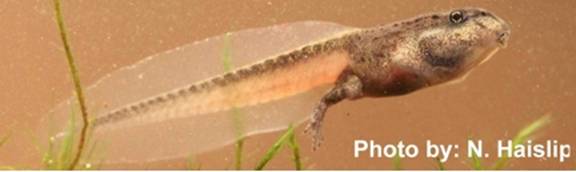
Nathan Haislip
M.S. Candidate

Research
Amphibian populations are declining
globally and one cause is emerging infectious diseases. Ranaviruses are
responsible for the majority of amphibian mass mortality events in North
America, yet research into factors that govern host susceptibility is limited. Ecological stressors may be important
components contributing to the emergence of infectious diseases, and two known
stressors are predation and development. In response to predators, prey may
adaptively alter their behavior, morphology, and life history traits. Although
enhancing survival, stress responses may also negatively impact immune
functions if they persist in an organism. Thus, a constant threat of predators
could increase the susceptibility of an organism to pathogen infection and
contribute to the emergence of infectious diseases. Evidence also exists
that there are varying degrees of immune system development across different
amphibian life stages and that susceptibility to ranaviruses may differ across
these life stages. Unfortunately,
studies comparing the susceptibility of amphibians to pathogens at different
developmental stages are rare. My research focuses on determining the
impacts of natural stressors on ranavirus
emergence.
The objectives of my
study are:
1) Quantify
the effects of amphibian development on ranavirus
pathogenicity, and
2) Quantify
the effects of predators on ranavirus
pathogenicity.
I am also performing surveillance
for ranavirus outbreaks in larval amphibian populations among 40 wetlands over
two years in the Cumberland Plateau and the Tennessee River Ridge and Valley
physiographic regions. Lastly, I am working in collaboration with Dr. Jason Hoverman. He
is comparing the relative susceptibility of amphibian species across multiple
families to novel verses endemic ranavirus strains.
Ranavirus Infection:
Through experimental
challenges at the JARTU
facility, we have documented numerous physiological changes that are
associated with ranavirus infection and morbidity. Gross signs included edema (swelling, Fig.
1), erythema (reddening, Fig. 1, 2), loss of pigmentation, and hemorrhages. Behavioral signs include problems with buoyancy,
lethargy, inappetence, and erratic swimming. Internal gross signs included
swollen organs, hemorrhaging of organs and fat bodies (Fig. 3), congested blood
vessels, and paleness of the organs (Fig. 3). Most internal signs were
associated with the kidney and liver, which is not surprising considering these
organs are known targets of ranaviral infection.
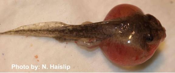
Figure 1: Edema and erythema
of Hyla chrysoscelis.
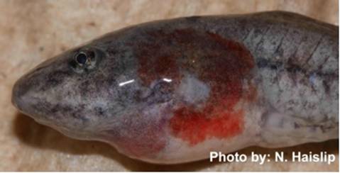
Figure 2: Erythema of Rana clamitans.
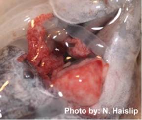
Figure 3: Hemorrhaging
of fatbodies and paleness of liver.
Collaborators:
Tennessee Agricultural Experiment Station, UGA
Veterinary Diagnostic and Investigational Laboratory, and Tennessee
Wildlife Resources Agency
Personal
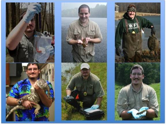
Hello!
I grew up in a
small town in middle Tennessee approximately 60 miles south of Nashville.
I graduated with honors from the University of Tennessee with a B.S. in Wildlife and
Fisheries Science, and was recruited into the UT Center for Wildlife
Health to conduct Ranavirus research. While I was an
undergraduate, I conducted two independent studies on grassland snake
populations, was an intern with the Tennessee Wildlife Resources Agency,
assisted with grassland songbird research in middle Tennessee, and worked for a
year with Dr. Gordon Burghardt with
multiple species of snakes and monitor lizards (above photo). I also was
very active in the UT student chapter of The Wildlife Society,
and volunteered on various projects. My passion is herpetology and
wildlife photography. I am also an avid birder, hunter, and fisherman.
Contact Information
Email: nhaislip@utk.edu
Phone: 865-974-3897
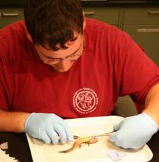
(Larval Ambystomatid
necropsy for Ranavirus testing)
UT Department of
Forestry, Wildlife and Fisheries